 Know Thy Body
Field guide/Plates · Cross-sections
Cut open
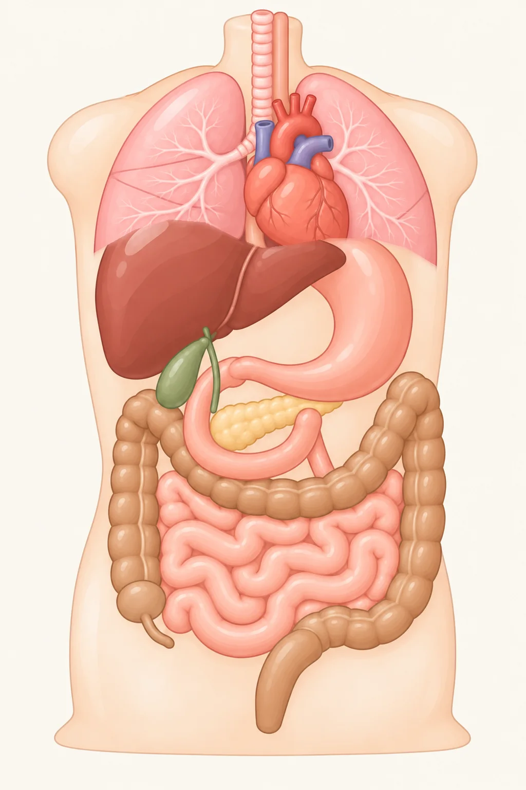
From the front, organs stack one behind another: the esophagus hides behind the windpipe, the pancreas hides behind the stomach, and the kidneys tuck behind the intestines. Only when you cut through do you see what sits in front and what sits in back – first split straight down the middle, then sliced across at six levels from the base of the neck to the pelvis.
- Proper name
- Transverse plane Planum transversale
- View
- From the feet looking up, like a CT scan
- Orientation
- Front at the top; body’s right on the left
Side view: split down the middle, front to back
Before slicing across, cut straight down the middle from head to pelvis and look from the body’s left side: front is on the left, back on the right. The whole front-to-back order shows up at once: heart behind breastbone, esophagus and aorta behind heart, spine behind those; below the diaphragm, liver and stomach sit in front with the pancreas tucked behind the stomach against the spine; down in the pelvis, bladder sits in front and rectum in back. Both lungs and both kidneys sit off to the sides, so a midline cut misses them. The six dashed lines mark where the horizontal slices cut across; tap any label on the map to jump straight to that level.
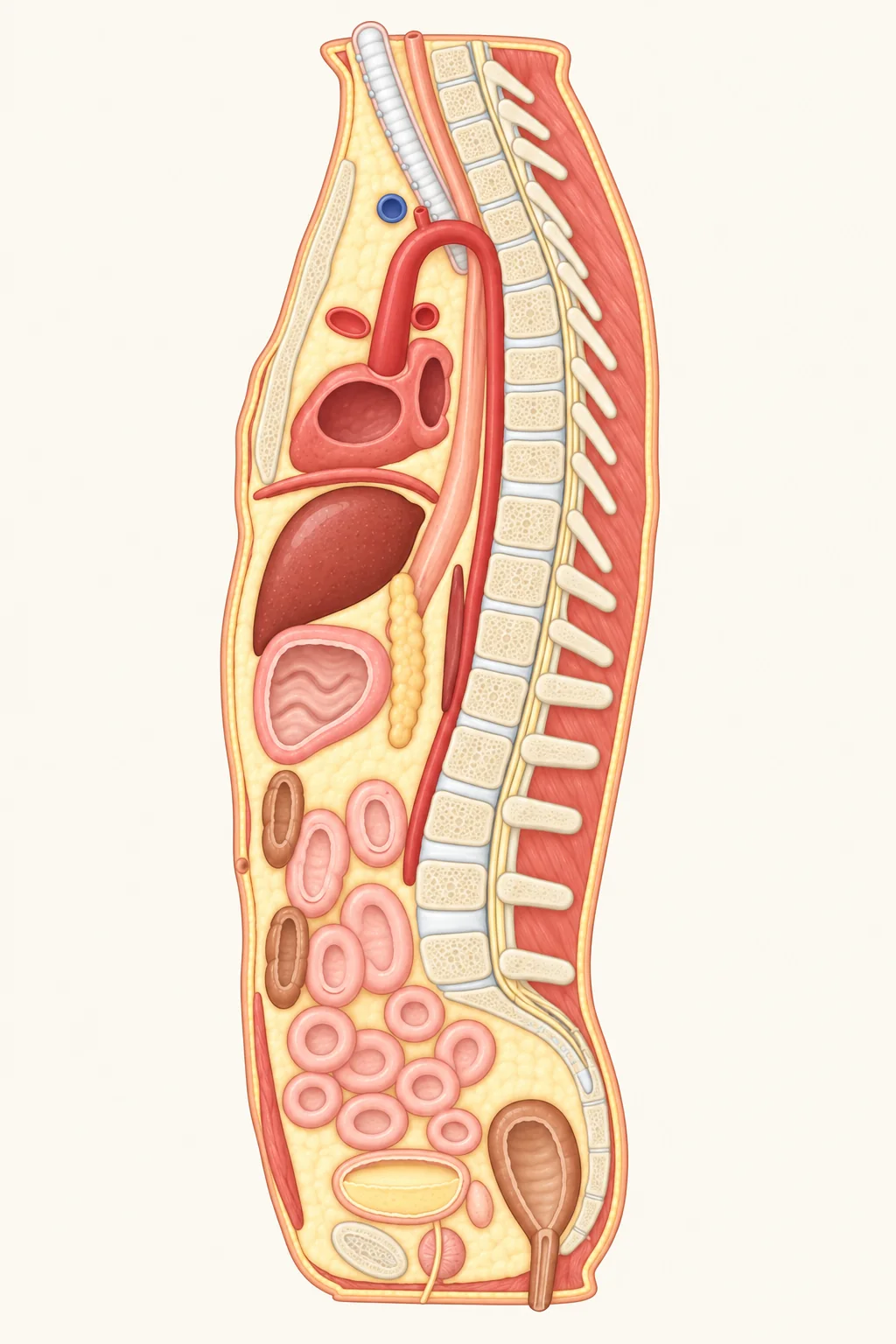
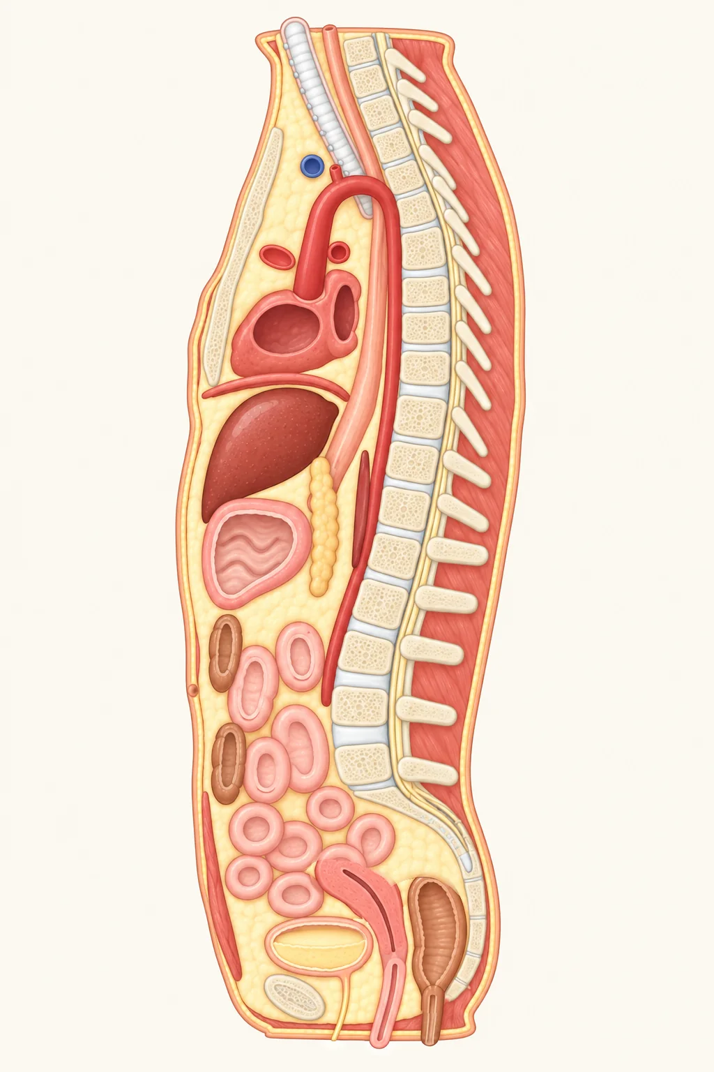
Tap an organ in the drawing or the list below to see why it sits there.
- WindpipeRuns down from the throat into the chest in front of the esophagus, then splits between the 4th and 5th thoracic vertebrae into left and right bronchi for each lung – so this midline cut catches only the stretch above the split. Know your lungs →
- EsophagusTravels down right between the windpipe and the spine, passes directly behind the heart, slips through the diaphragm, and joins the stomach just below it. Know your esophagus →
- BreastboneThe flat bone down the center of your chest, made of the manubrium, body, and xiphoid tip. Your heart rests directly behind it – during CPR, chest compressions push on its lower half.
- HeartSits behind the breastbone, tilted mostly toward the left; rests on the diaphragm below and presses against the esophagus behind through its sac. Know your heart →
- AortaRises from the heart, arches back and to the left behind the top of the breastbone, then runs down hug-tight against the spine through the diaphragm into the belly before splitting near L4 into arteries for each leg.
- DiaphragmA dome of muscle separating chest and belly like an upside-down bowl: the heart sits on top of the dome, and the liver presses against its underside. When you breathe in, it contracts and flattens downward, pulling air into the lungs.
- LiverTucks straight under the diaphragm, separated from the heart above only by the diaphragm and heart sac. Most of it fills your right upper belly; this midline cut clips the wedge reaching toward the center. Know your liver →
- StomachSits below the liver toward the front belly wall, with the pancreas tucked directly behind it. Know your stomach →
- PancreasSide view makes it obvious how deep it hides: behind the stomach in front, and right in front of the aorta and spine in back – which is why acute pancreatitis pain often bores straight through to the back. Know your pancreas →
- DuodenumThe first segment of the small intestine, curving into a C-shape around the head of the pancreas. Its bottom stretch crosses horizontally in front of the aorta, so a midline cut slices it into an oval ring. Know your small intestine →
- ColonThe upper circle here is the transverse colon draping below the stomach; the lower circle is the sigmoid colon leading into the rectum. Know your large intestine →
- Small intestineCoils back and forth through the middle of the belly all the way down into the pelvis above the bladder, so every loop cut across shows up as a ring. Know your small intestine →
- Small intestineCoils back and forth through the middle of the belly all the way down into the pelvis above the uterus, so every loop cut across shows up as a ring. Know your small intestine →
- Belly buttonWhere the umbilical cord attached before birth; once cut, the stump dried and fell off, leaving a small scar roughly level with the disc between L3 and L4.
- Abdominal muscleTwo long strap muscles (rectus abdominis – the “six-pack” muscles) running down the front of your belly from ribs to pubic bone. A dead-center cut follows the fibrous seam between them, catching only the lower edge.
- BladderSits all the way at the bottom right behind the pubic symphysis. Empty, it stays hidden inside the pelvis; as it fills with pee, it domes upward above the pubic bone. Know your bladder →
- Pubic symphysisThe cartilage-padded joint where the left and right pubic bones meet in the front midline – the firm ridge you can feel right at the bottom of your lower belly.
- ProstateSits directly beneath the bladder, wrapping like a ring around the top stretch of the urethra – so when it enlarges, it squeezes the outlet and slows your stream or wakes you up at night.
- Seminal vesiclesA pair of glands tucked behind the bladder between bladder and rectum, producing roughly 60% of semen’s fluid.
- UrethraExits the bottom of the bladder and passes straight through the prostate; in males it runs about 8 inches (20 cm) total. Know your bladder →
- UterusSits between the bladder and rectum, usually leaning forward over the top of the bladder; as the bladder fills, it tilts the uterus upright. Its lower neck (cervix) opens into the vagina. Late in pregnancy, a growing uterus and baby’s head press directly on the bladder, sending you to the bathroom often.
- VaginaAn elastic muscular canal running from the cervix down to the outside, sitting behind the bladder and urethra and in front of the rectum.
- UrethraExits the bottom of the bladder and runs only about 1.2–1.6 inches (3–4 cm) to open just in front of the vaginal opening. Know your bladder →
- RectumFollows the curve of the sacrum and tailbone downward into the anal canal. Know your large intestine →
- SpineFrom the side, your spine has four gentle curves: neck and lower back curve forward, while mid-back and sacrum curve back. Lumbar vertebrae carry the most body weight, making them the thickest blocks.
- Spinal discCushions sitting between vertebrae: tough fibrous rings outside around a gel-like center. A slipped (herniated) disc happens when that soft center bulges backward through weakened rings and presses on nearby nerves – most often between L4–L5 or L5–S1.
- Spinal cordRuns down inside the spinal canal, ending in adults around the first or second lumbar vertebra (L1–L2). Below that lies only a loose bundle of nerve roots (the cauda equina), which is why a lumbar puncture in the lower back misses the spinal cord safely.
Base of the neck: windpipe in front, esophagus behind
This cut crosses where your neck meets your chest: the little notch you can feel at the very top of your breastbone lines up roughly with the lower edge of the 2nd thoracic vertebra (T2). Even up here at the base of the neck, the tops of both lungs poke into view. Between the lungs, from breastbone to spine, lies the mediastinum – a tight corridor carrying the windpipe, the esophagus, and great vessels heading to and from your head and arms.
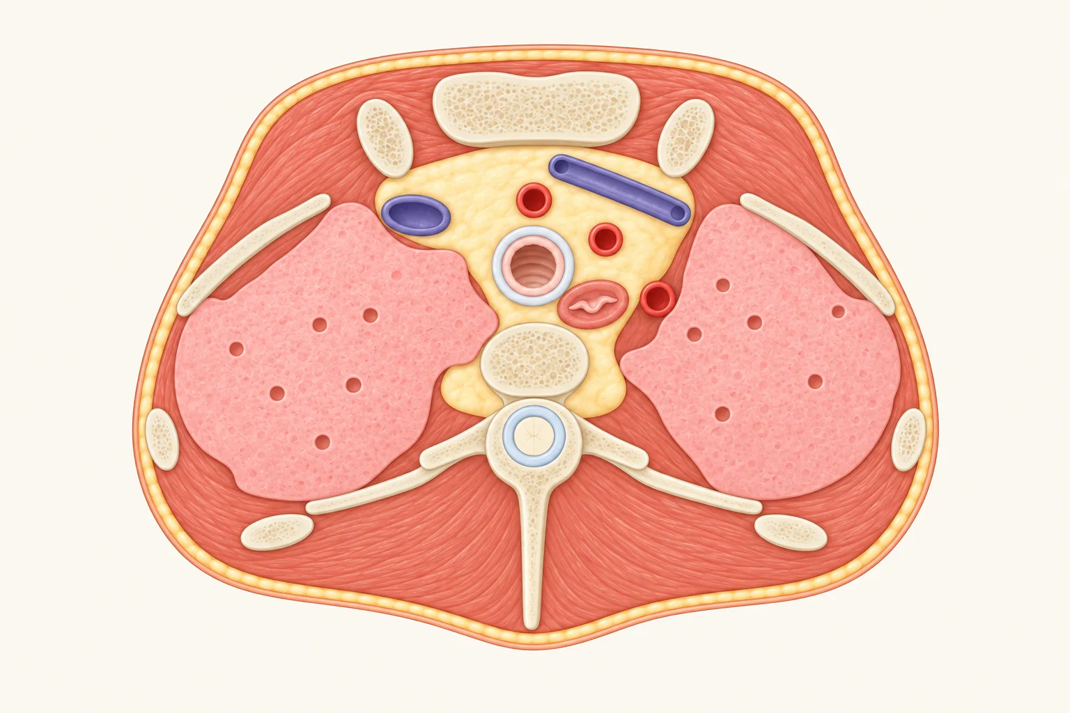
Tap a structure on the slice or in the key below to see why it sits there.
- LungsHere you’re seeing the very tops (apices) of both lungs. Lungs rise higher than most people think – peeking about 1–1.5 inches (2.5–4 cm) above the front of the first rib, tucked just above the inner end of each collarbone. Know your lungs →
- WindpipeDead center and toward the front. Stacked from 16 to 20 C-shaped cartilage rings so it never collapses; the open back of each C is bridged by flexible smooth muscle pressing right against the esophagus. Know your lungs →
- EsophagusTucked straight behind the windpipe and in front of the spine, drifting slightly left as it drops from the neck before centering around T5. Know your esophagus →
- Brachiocephalic veinsTwo veins draining blood back from your head, neck, and arms. The left one runs roughly 2.4 inches (6 cm) obliquely behind the breastbone toward the right; the right one is only about 1 inch (2.5 cm) long. Behind the first right rib cartilage they merge into the superior vena cava.
- ArteriesThree major branches rising off the aortic arch. On the right, a single brachiocephalic trunk divides into the right common carotid and right subclavian arteries; on the left, both the left common carotid and left subclavian spring directly from the arch, feeding head, neck, and arms.
- BreastboneThis top block is the manubrium. Its upper notch sits roughly level with the bottom of T2 (slightly lower in women, closer to T3).
- CollarboneIts inner end meets the top of the breastbone here at the sternoclavicular joint – the only bone-to-bone joint connecting your entire arm to your trunk.
- RibsThe first rib is short and sharply curved, looping around the lung apex into a long diagonal strip here; the ovals along the sides and back are downward-slanting 2nd and 3rd ribs caught mid-slice.
- Thoracic vertebraT2. The round hole in the middle is the spinal canal carrying the spinal cord; the prong sticking straight back is the spinous process – part of the bony bumps you feel down the center of your back.
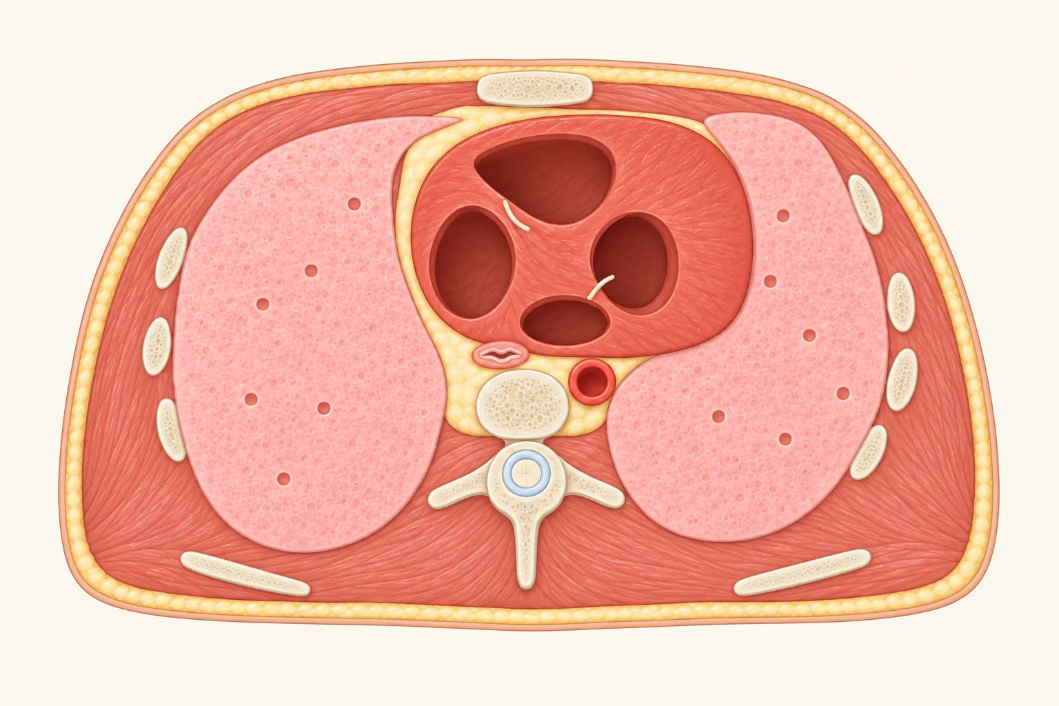
Heart level: all four chambers in one slice
Drop down to the 8th thoracic vertebra (T8), and the cut runs straight through all four chambers of the heart: the right ventricle takes up most of the front right behind the breastbone; the thick-walled left ventricle sits to the left; and the left atrium sits furthest back. Because the heart leans left, the left lung notches inward to make room, leaving it slightly smaller than the right.
vertebraFront · chestBackRL
Tap a structure on the slice or in the key below to see why it sits there.
- HeartRoughly two-thirds sits left of the midline. All four chambers appear here: the right ventricle fills most of the front behind breastbone and rib cartilage; the left ventricle on the left has walls about three times thicker to pump blood around the body; the left atrium sits at the back, and the right atrium on the right. Know your heart →
- LungsCradling the heart on both sides. The left lung holds slightly less volume and carries a hollow curve (the cardiac notch) for the heart, while the right lung is wider and a little shorter. Know your lungs →
- EsophagusHugs right behind the heart, pressing up against the left atrium across the heart sac. That close fit is why doctors slide a thin ultrasound probe down the esophagus (transesophageal echo) for crystal-clear heart images with no ribs or lung air in the way. Know your esophagus →
- AortaHere the descending thoracic aorta runs downward just left-front of the spine, shifting steadily toward the midline as it nears the diaphragm.
- BreastboneYour heart sits right behind it – hands go over the lower half of this bone in the center of the chest during CPR.
- RibsEach oval ring on either side is a slanting rib caught across the cut. Twelve pairs join twelve thoracic vertebrae in back and the breastbone in front to cage your heart and lungs.
- Shoulder bladeTwo flat triangular bones floating across the upper back, held in place not by joints with the ribs, but entirely by surrounding muscles.
- Thoracic vertebraT8, with the spinal cord passing down through the central spinal canal.
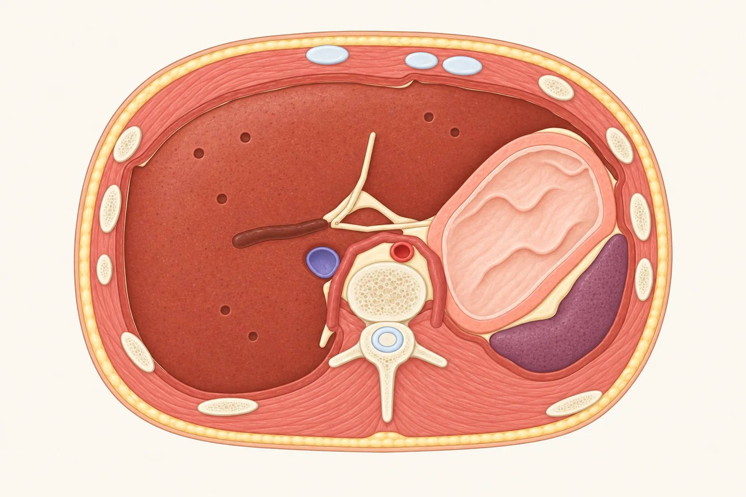
Below the diaphragm: liver and stomach tucked behind ribs
Slice down at the 11th thoracic vertebra (T11), and the lungs have disappeared. Think of the diaphragm as an upside-down dome: heart and lungs sit above the roof, while liver, stomach, and spleen tuck underneath. Even though this cut already lies below the dome inside the belly, the lower ribs still wrap all the way around – sheltering the liver and spleen behind bone.
Tap a structure on the slice or in the key below to see why it sits there.
- DiaphragmA dome of muscle dividing chest and belly and your main breathing muscle – contracting and flattening downward to pull air in. Two muscular legs (crura) anchor it to the upper lumbar spine, arching right over the aorta so the vessel passes underneath.
- LiverYour largest internal gland, filling most of the upper right belly right beneath the diaphragm and safely tucked behind your right lower ribs. Know your liver →
- StomachIts upper portion appears here on the left toward the front, resting alongside the left side of the liver with the spleen behind it. Know your stomach →
- SpleenTucked up in the far upper left belly behind the stomach and under the diaphragm, shielded behind the left 9th to 11th ribs.
- Inferior vena cavaNestled into a groove along the back of the liver before piercing the diaphragm (around T8–T9) and entering the right atrium roughly 1 inch (2.5 cm) higher, returning blood from your lower body.
- AortaSitting straight in front of the spine, framed between the diaphragm’s two muscular legs before slipping under that arch into the belly at T12.
- RibsThe lower ribs still curve around both sides at this height, shielding the liver on the right and the spleen on the left.
- Thoracic vertebraT11 – next-to-last of the twelve thoracic vertebrae.
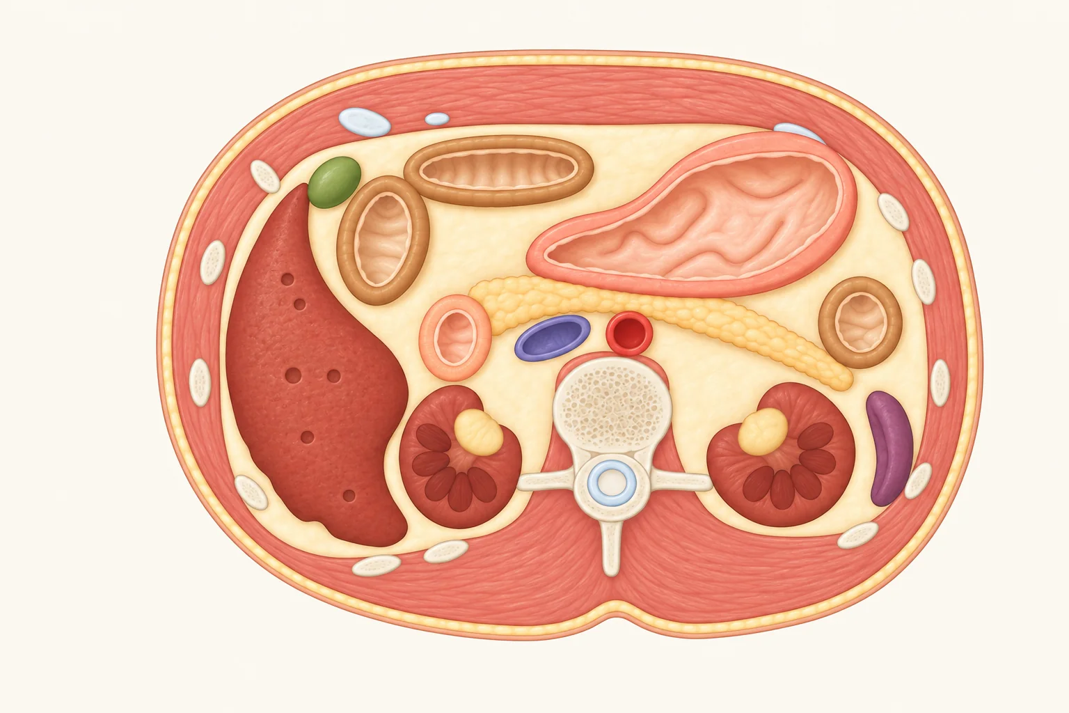
First lumbar vertebra (L1): pancreas and kidneys sit here
Roughly halfway between the bottom of the breastbone and your belly button sits the 1st lumbar vertebra (L1), known in anatomy as the transpyloric plane: the stomach’s exit (pylorus), the pancreas, both kidney entryways, two sharp bends of the colon, and usually the tip of the gallbladder all line up right around this height.
Tap an organ on the slice or in the key below to see why it sits there.
- LiverStill occupies much of the upper right belly from front to back, mostly tucked under the lower right ribs. Know your liver →
- StomachRests in the upper left front belly right against the abdominal wall, expanding after a meal and shrinking when empty so its outline looks different from person to person and hour to hour. Know your stomach →
- PancreasLies flat directly behind the stomach: its head cradled in the duodenum on the right, its tail stretching left toward the spleen – hidden behind the stomach from a front view. Know your pancreas →
- KidneysBoth sit all the way in the back beside the spine, tucked behind the liver, stomach, pancreas, and gut – which is why kidney pain feels deep in your flank under the ribs. Know your kidneys →
- GallbladderTucked under the liver near the front abdominal wall, its rounded tip lying roughly beneath the tip of the 9th right rib cartilage right around this slice. Know your gallbladder →
- DuodenumThe first section of the small intestine leaving the stomach and curving into a C-shape around the head of the pancreas. Know your small intestine →
- ColonFrames the belly like a square border; three stretches of it get caught in this cross-section. Know your large intestine →
- SpleenTucked far to the outer left back under the left ribs, filtering out worn red blood cells and helping fight infection.
- AortaThe body’s largest artery descending straight in front of the spine, slightly left of center.
- Inferior vena cavaThe main vein returning lower-body blood to the heart, running parallel right beside the aorta on the spine’s front-right side.
- Lumbar vertebraL1, one block in the spinal column, with the spinal cord passing through its central canal.
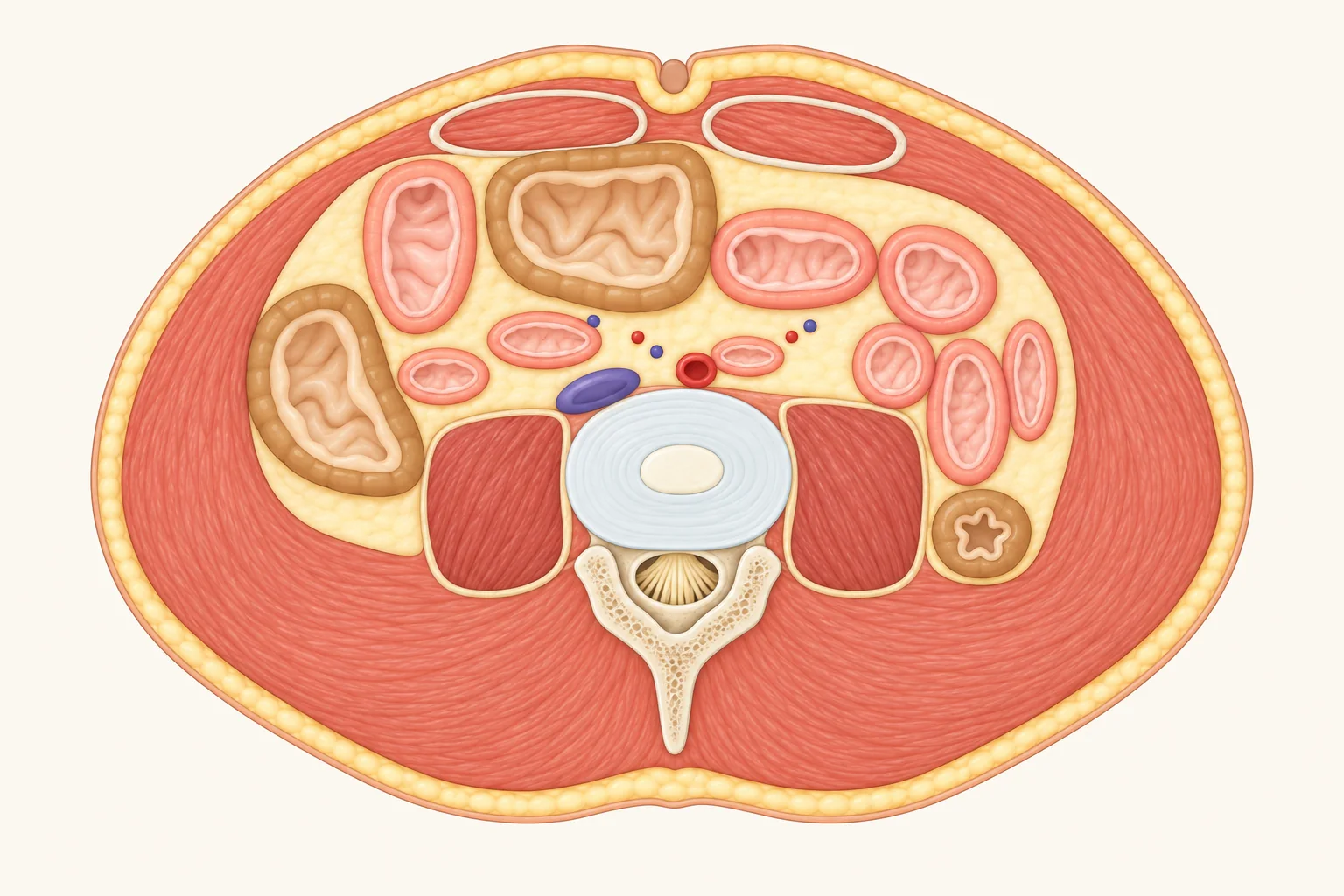
Navel level: small intestine coiled in the middle
Your belly button sits roughly level with the disc between the 3rd and 4th lumbar vertebrae (L3–L4). Below both kidneys and still above the pelvis, the colon frames the outer edges while the small intestine loops back and forth across the middle, with the aorta and inferior vena cava resting side by side against the spine.
colonTransverse
colonDescending colonSmall
intestinePsoasDiscAortaInferior
vena cavaAbdominal
muscleBelly
buttonCauda equinaLumbar vertebraFront · navelBack · lower backRL
Tap a structure on the slice or in the key below to see why it sits there.
- Small intestineAbout 10 feet (3 m) long in a living adult, coiled back and forth across the middle belly so every loop looks like a circle in cross-section. Inside, circular folds, villi, and microscopic microvilli multiply its absorbing surface area more than 600-fold. Know your small intestine →
- ColonAscending colon on the right, transverse colon dipping across the front, and narrower descending colon on the left – framing the small intestine inside. Know your large intestine →
- Spinal discThe shock-absorbing pad between lumbar vertebrae: tough fibrous rings outside around a soft gel core. When that core bulges backward into nerves, it causes a herniated disc – most often at L4–L5 or L5–S1, just below this slice.
- Cauda equinaSince the spinal cord ends around L1–L2, the canal at this lower height holds only a dangling bundle of nerve roots shaped like a horse’s tail (cauda equina) – so a lower-back spinal tap avoids the spinal cord itself.
- PsoasA thick pair of muscles hugging either side of the lumbar spine from T12–L5 down to the thigh bone, working with the iliacus muscle to flex the hip and lift your leg forward.
- AortaSitting just left of center in front of the spinal disc – a bit lower around L4 it splits into left and right iliac arteries heading into the legs.
- Inferior vena cavaRight beside the aorta, carrying blood upward from both legs and the lower body back toward the heart.
- Abdominal muscleTwo parallel strap muscles (“six-pack” muscles) in front, segmented by cross-bands and joined down the center by a fibrous seam right where the belly button sits.
- Belly buttonThe healed scar left after the umbilical cord dried and fell off after birth, typically lining up near the L3–L4 disc.
- Lumbar vertebraBecause this cut slices through the disc between two vertebrae, the front shows the disc while the back clips the bony arch guarding the spinal canal and its rearward prong.
Pelvis: bladder in front, rectum behind
The lowest slice cuts right across the hip joints, slicing cleanly through the ball-shaped top of each thigh bone. Inside the bowl of the pelvis, both males and females have the bladder in front and the rectum in back; what differs is what sits in between – seminal vesicles in males, uterus in females.
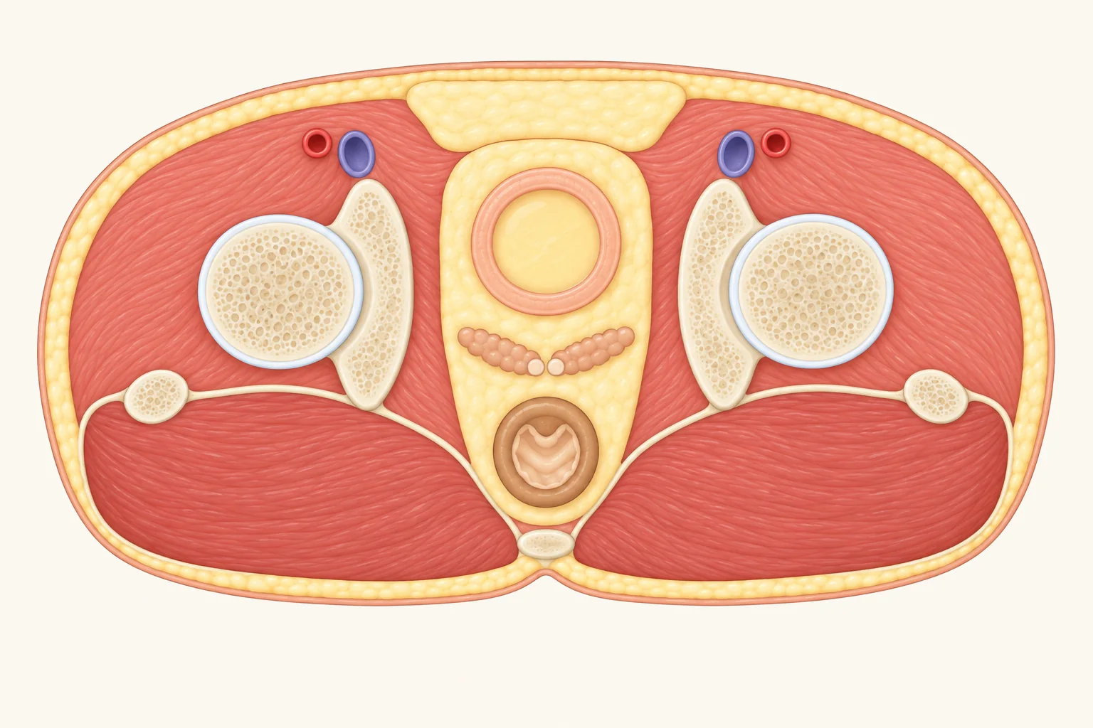
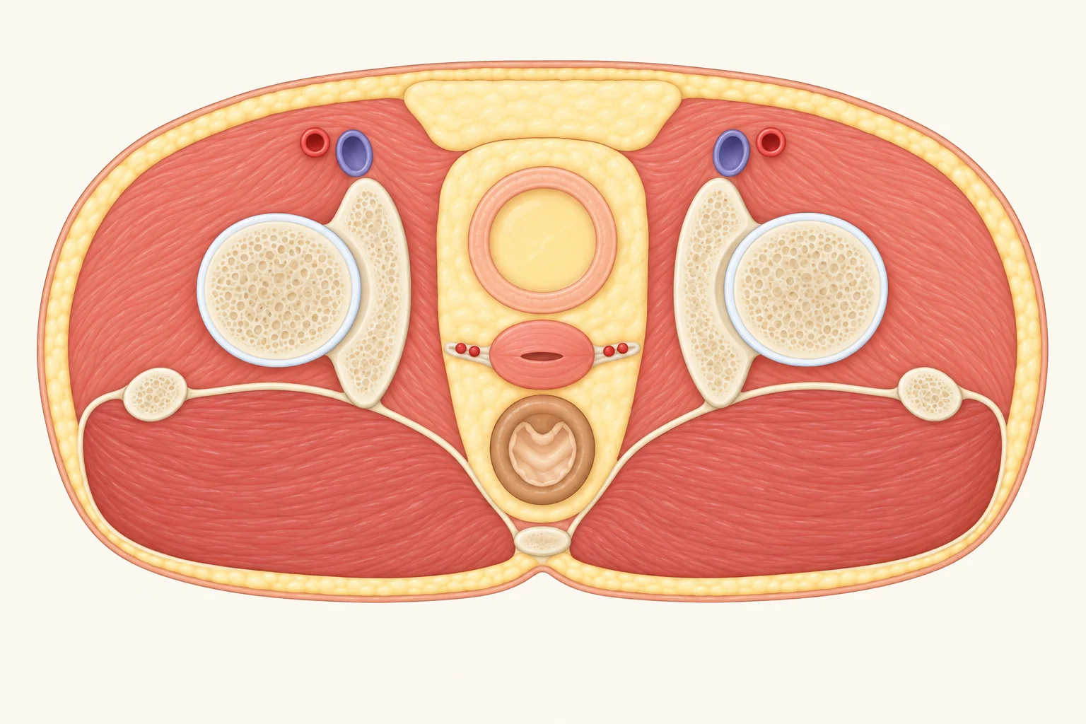
Tap a structure on the slice or in the key below to see why it sits there.
- BladderTucked right at the front directly behind the pubic bone. Know your bladder →
- Seminal vesiclesA pair of glands nestled right behind the bladder between bladder and rectum, making about 60% of semen’s fluid. The prostate sits slightly lower beneath the bladder, just under this slice.
- UterusSits right between the bladder in front and the rectum in back. Late in pregnancy, a growing uterus and baby’s head press directly down on the bladder, explaining frequent bathroom trips.
- RectumSits all the way in the back against the front face of the sacrum and tailbone. Know your large intestine →
- FemurThe rounded ball (femoral head) at the top of the thigh bone snaps into the hip bone’s deep socket to form a ball-and-socket joint that lets your leg swing in any direction; the smaller rounded bump to the outside is the greater trochanter.
- Hip boneForms the sides of the pelvis, cradling the head of the thigh bone inside its cup-shaped socket (acetabulum).
- Femoral vesselsThe femoral artery and vein run down the front of the upper thigh right below the crease of the groin, roughly halfway between the front hip point and the pubic midline – where you can feel a strong groin pulse.
- Buttock muscleThe gluteus maximus – largest of the three gluteal muscles and one of the biggest, strongest muscles in the whole body, extending the thigh backward and pulling your torso upright when you stand from bending over.
- TailboneThe very tip at the bottom of your spine, usually three to five (most often four) tiny fused vertebrae.
How to read a cross-section
Picture someone lying flat on their back while you stand at their feet looking up toward their head: front (belly) sits at the top of the slice, back at the bottom, and their right side sits on your left. Hospital CT and MRI scans are oriented the exact same way, so these drawings match medical scans.
The midline side view works differently: imagine standing beside the person’s left hip looking at their profile – front on the left, back on the right, and head at the top.
If anything on a slice looks crowded, tap or hover over it to light it up – or tap any row in the key (or navigate with your keyboard) to highlight where it sits.